Left axillary a Left vertebral a Left carotid sinus Right subclavian a. Veins and arteries Labeling review Head Neck Lab Quiz Review.
Solved Label The Arteries Of The Neck In The Ct Angiogram Chegg Com
From 1995 to 1998 16 patients with suspected traumatic carotid artery injury underwent CTA.
. This is one of the safest ways to study the head and neck. A CT scan can reduce or avoid the need for invasive procedures to diagnose problems in the skull. MRI of the head.
Computerized tomographic angiography also called CT angiography CTA is a radiological test that combines the technology of a conventional CT scan with that of traditional angiography to create detailed images of the blood vessels in the body. Label the arteries of the aortic arch in the CT angiogram. A CT coronary angiogram uses a powerful X-ray machine to produce images of your heart and its blood vessels.
Other tests that may be done instead of CT scan of the head include. A CT scan can reduce or avoid the need for invasive procedures to diagnose problems in the skull. While CT angiography should not be used as a screening test in the general population it is a major new tool in the diagnosis of coronary artery diseaseIn patients at high risk for developing coronary disease cigarette smokers those with genetic risk high cholesterol levels hypertension or diabetes who have unclear results with treadmill or other testing or who have symptoms.
Right common carotid a. This retrospective study was done to evaluate the use of CT angiography CTA in suspected vascular injuries of the neck. An angiogram of the neck carotid angiogram can be used to look at the large arteries in the neck that lead to the brain.
Terms in this set 32 Internal jugular vein. An angiogram of the head and neck is an X-ray test that uses a special dye and imaging fluoroscopy to take pictures of the blood flow in the blood vessels of the head and neck. CT angiography image of an anterior communicating artery aneurysm in the brain arrow.
CT angiography angio of the neck or brain is a specialized study of the arterial anatomy of the respective region. CT angiography of the cerebral arteries also known as a CTA carotids or an arch to vertex angiogram is a noninvasive technique allows visualization of the internal and external carotid arteries and vertebral arteries and can include just the intracranial compartment or also extend down to the arch of the aorta. Left internal carotid a.
Left internal carotid a. CT angiography CTA evaluates the major vessels of the head neck or both. Label the blood vessels and structures using the hints provided.
Mid segment of the left anterior descending LAD artery small first diagonal artery D1 small ramus intermedius artery RI first septal artery S1 left coronary sinus of the aortic valve. The overarching goal of this examination is an optimal. It is not a particularly original work and you will easily find hundreds of cross-sectional atlases out there.
Anatomy and Physiology questions and answers. Twelve of these patients had penetrating injuries and four had blunt injuries to the neck. A computerized tomography CT coronary angiogram is an imaging test that looks at the arteries that supply blood to your heart.
CT scan of head and neck. Second diagonal artery D2 right coronary sinus of the aortic. Where it stands out I think is in the detail of vessel labeling.
CT scan of head and neck. An angiogram of the neck carotid angiogram can be used to look at the large arteries in the neck that lead to the brain. CT scan of head and neck.
This is one of the safest ways to study the head and neck. Label the specific arteries taking blood to the head and neck region. While dedicated CT angiography is generally superior for the specific evaluation of arterial disease many arterial abnormalities can be identified on standard soft-tissue neck CT images.
The vertebral arteries provide blood flow to the back of the brain A tear in the wall of an artery dissection. An angiogram of the head and neck is an X-ray test that uses a special dye and camera fluoroscopy to take pictures of the blood flow in the blood vessels of the head and neck. Radiological anatomy of the head and neck on a CT in axial coronal and sagittal sections and on a 3D images.
Other tests that may be done instead of CT scan of the head include. All Veins and Arteries are in singular form this will be easier for the test I also removed left and right. CTA can assess the status of both the large arteries and veins in most parts of the body including.
A CT angiogram of an elderly patient obtained for evaluation of carotid stenosis has been labeled in regard to both vessels and adjacent structures. Left external carotid a Left subclavian a. CT angiography image of the head and neck showing the major arteries.
Left axillary a Left vertebral a Left carotid sinus Right subclavian a. An iodine based contrast agent is rapidly injected through an IV placed in a vein usually in the arm. An angiogram of the neck carotid angiogram can be used to look at the large arteries in the neck that lead to the brain.
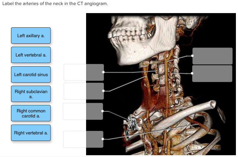
CT scan of head and neck. Label the arteries of the neck in the CT angiogram. Arch of aorta Brachiocephalic trunk Left common carotid a.
MRI of the head. Label the arteries of the neck in the CT angiogram. Being exposed to radiation.
Label the arteries of the neck in the CT angiogram. An aneurysm is a bulge in the artery wall which can in certain circumstances rupture. Narrowed or blocked vertebral artery in the neck.
Label the blood vessels using the hints provided. The data can then be reviewed in multiple planes and 3 dimensional images can also be created. Right vertebral a.
The procedure is noninvasive and doesnt. An angiogram of the head cerebral angiogram can be used to look at. Subclavian artery brachiocephalie trunk external carotid artery common carotid artery internal carotid artery 2.
Identification of arteries and veins 1. An angiogram of the head and neck is an X-ray test that uses a special dye and camera fluoroscopy to take pictures of the blood flow in the blood vessels of the head and neck. Risks Risks for CT scans include.
Label the arteries of the neck in the CT angiogram. Positron emission tomography PET scan of the head. The common carotid internal carotid and vertebral arteries traverse the neck and can be readily evaluated at standard contrast-enhanced neck CT.
A weak area in the wall of a blood vessel that causes the blood vessel to bulge or balloon out aneurysm. Positron emission tomography PET. It might be done to diagnose the cause of chest pain or other symptoms.
A CT scan uses x-rays to acquired images as the contrast bolus passes through the arteries.

Lab 7 Blood Vessels Flashcards Quizlet

Pdf Blood Vessel Segmentation For Neck And Head Computed Tomography Angiography Semantic Scholar

Solved Label The Arteries Of The Neck In The Ct Angiogram Chegg Com

0 Comments